Plastic embedding
Cells are typically
- fixed with 2% glutaraldehyde (e.g. in 100 mM Na-cacodylate or Na-phosphate buffer, pH 7.4) for 1-2 hours at RT
- post-fixed with 1% osmiumtetroxide for 1 h at RT
- dehydrated with graded series of ethanol (70%, 96%, 100%)
- incubated with transitional solvent acetone
- embedded gradually in Epon
So-called flat embedding technique is suitable for cell monolayers cultured on class coverslips. In this technique, processing is done while cells are still attached to the coverslip, and the coverslip is removed only after polymerization assuring well preserved morphology. Even individual cells can be processed when grown on Cellocate-coverslips (Eppendorf) or Glass bottom culture dishes with grid (MatTek).
Most commonly used fixatives are Glutaraldehyde (50% stock solution, EM-grade, Fluka) and Paraformaldehyde (16% solution in 10 ml vials, EM-grade, Electron Microscopy Sciences, EMS).Osmiumtetroxide is from EMS. Resins used at our unit are Epon (TAAB Embedding resin, medium), Low viscosity resin (Agar; replacing Spurr-resin, which is not available anymore) and Lowicryl HM20 (TAAB).
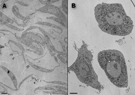
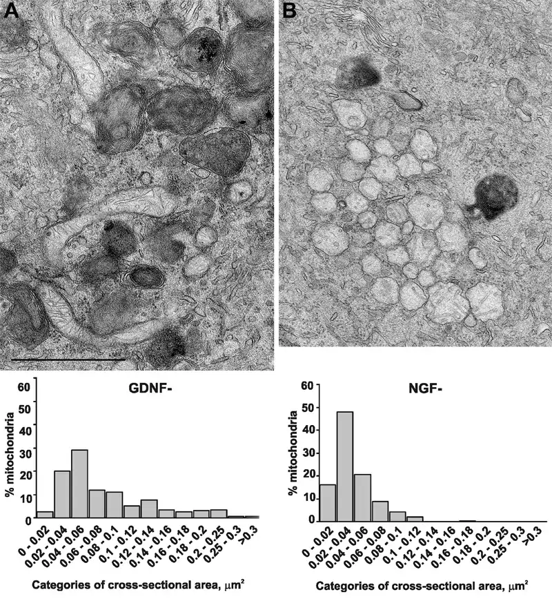
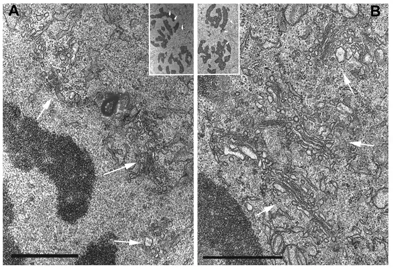
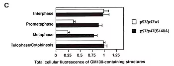
Image 1 Cell monolayers can be sectioned parallel or perpendicular to the coverslip. NRK-52E cells were either detached from the substratum and processed as cell pellet (A) or grown on glass coverslip and flat embedded (B and C). In B, the sections were cut parallel to the coverslip, whereas in C, sectioning was perpendicular to the coverslip (E. Jokitalo).
Image 2 Mitochondria of NGF-deprived (A), but not GDNF-deprived (B) sympathetic neurons are structurally changed. The morphological change is analyzed quantitatively by determining the cross-sectional areas of the mitochondria in images taken from both specimens. (Yu et al., 2003 JCB 163:987-997)
Image 3 and 4 The microinjection of p97/p47(S140A) preserves Golgi stacks (B) in mitotic late prometaphase cells whereas wtp97/p47 injected cells have typical mitotic Golgi clusters (A). Table (C)shows percentage of total Golgi membranes in each three categories (Uchiyama et al., 2003 JCB 161:1067-1079).